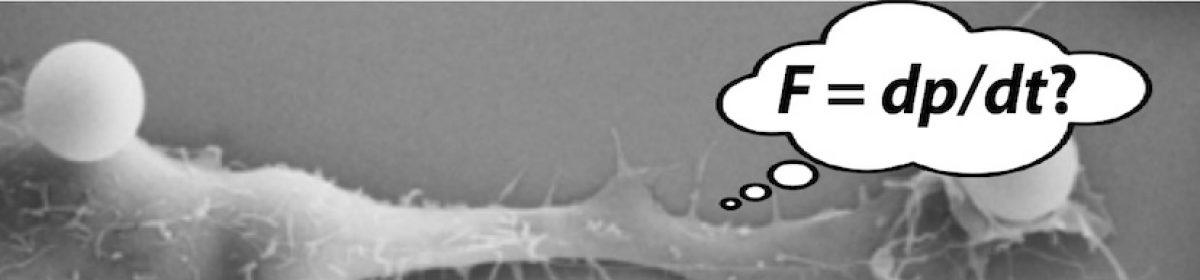
Motivation
All of these types of motion can be observed in onion cells. Therefore, onion cells are a cheap and simple experimental subject that allows us to make several interesting observations. By looking at individual onion cells, we can make both qualitative and quantitative observations about the different types of motion. Moreover, analysis of the work required to move the vesicle, based on terminal speed and energy required for ATP hydrolysis (23 kJ/mole), can give us insight into how fast ATP is utilized during these transport processes.
Materials and Methods
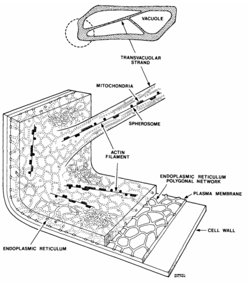
In order to observe motion inside of individual onion cells, we must first prepare a slide with one layer of onion cells. Fortunately, this is somewhat easy to do. To do this, cut down to the center of the onion. Activity in the onion cells is dependent on distance from the surface of the onion, and the center seems to be more active. Once you have a layer of onion close to the center, peel the lower cell membrane off of it. The cell membrane is relatively strong, and is made up of a single layer of cells. Put a few drops of saline solution down on a slide, place your onion membrane down, and then put a few more drops of saline solution down. Cover it with a slide cover, and blot the remaining saline solution. Keep in mind that onions are actually alive, but when you cut into it and mount the membrane on a slide, the cells will slowly die. The lifespan of cells in these slides is around 30 minutes. You might not observe cell activity on the first try; sometimes it will be necessary to try another section of the onion, or even another onion.
Slide prep
- Gently pull off the inner membrane from a section of onion. Anecdotally, the most active cells seem to come from an inner membrane from near the center of the onion.
- Clean a slide using a kimwipe; use a drop of saline if necessary.
- Put a drop of saline on your clean slide.
- Place your membrane on the drop of water; spread it out in a single layer and trim any ends that dangle over the edge of the slide.
- Put another drop of saline on top of the membrane.
- Drop a cover slip on top of your sample. Press it down gently to expel excess water.
- Blot extra water using a Kimwipe.
- Look at your sample under the highest magnification. The vesicles are small: about 1 um in size. If you don’t see any motion, move to a new location on the slide.
- I seem to get an active sample about 1 out of 3 preps. If you don’t see any motion after checking a dozen spots on your prep, try with a new one. You can throw away the cover slip but please re-use the microscope slide.
- Record your sample. You only need a minute or so of video.
- Use your prep reasonably promptly. They last 20 minutes or so.
Part A (week 1)
Take a video or two (be sure to note and record the average frame rate for the video) that show both apparently random and apparently directed motion. Track both types of motion using ImageJ and quantify the random and directed motion using the techniques we have developed in lab. Since you will be comparing these motions, when doing your analysis choose vesicles that are the same size.
Questions
- What is the diffusion coefficient for random motion of a vesicle?
- What is the speed of a vesicle undergoing active transport?
Part B (week 2)
You will use last week’s measurements of the diffusion coefficient of vesicles (from random motion) and the transport speed of vesicles (from directed motion) to estimate the viscosity of the cytoplasm and the rate at which ATP is burned during the transport process.
Writeup
Please use this blank template as a starting point for your writeup.
Diffusion questions
- What is the diffusion coefficient D of a typical vesicle?
- From D, what is the drag coefficient γ of a typical vesicle?
- Estimate the size of your vesicle. What is the effective viscosity of the interior of the cell?
Transport questions
Vesicles are transported by molecular motors (either myosin or kinesin) that consume ATP. Assuming that hydrolysis of 1 mole of ATP yields 23 kJ of energy under physiological conditions, and that molecular motors are 60% energy efficient, you can estimate how many ATP/sec are used in transporting a single vesicle.
- How much power (in Watts) is used in transporting a single vesicle at your measured speed v? Hint: you will need to use your value for γ from the previous question.
- This power is supplied by the hydrolysis of ATP in the cell. By considering the power balance between viscous drag and ATP-fueled transport, calculate how many ATP per second are consumed in transporting your vesicle.
Emergency data
Our onions were recalcitrant this year. Groups that didn’t get vesicle data, or whose data was low quality, may want to borrow some of this data from last year. I’m dumping all my files here: you will have to pick the one (or two) that works best for you. Use your calibration for 40x (maximum) magnification; the video was recorded at 6 fps.
https://ipls.gatech.edu/wp-content/uploads/2017/11/granules.wmv
https://ipls.gatech.edu/wp-content/uploads/2017/11/granules-bis.wmv
https://ipls.gatech.edu/wp-content/uploads/2017/11/granules-tris.wmv
https://ipls.gatech.edu/wp-content/uploads/2017/11/granules-quad.wmv
https://ipls.gatech.edu/wp-content/uploads/2017/11/granules-quint.wmv
https://ipls.gatech.edu/wp-content/uploads/2017/11/granules-sext.wmv
https://ipls.gatech.edu/wp-content/uploads/2017/11/granules-sept.wmv
https://ipls.gatech.edu/wp-content/uploads/2017/11/granules-oct.wmv
https://ipls.gatech.edu/wp-content/uploads/2017/11/granules-non.wmv